Figure 2
Neutralizing scFv Antibodies against Infectious Bursal Disease Virus Isolated From a Nlpa-Based Bacterial Display Library
Tianhe Li*, Bing Zhou*, Tingqiao Yu, Ning Li, Xiaochen Guo, Tianyuan Zhang, Jingzhuang Zhao, Liming Xu, Siming Li, Lei Ma, Tingting Li, Liangjun Ding, Mingzhe Sun, Deshan Li and Jiechao Yin
Published: 21 February, 2017 | Volume 1 - Issue 1 | Pages: 001-011

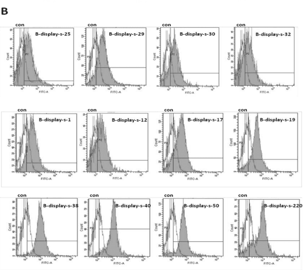
Figure 2:
Twelve VP2-binding scFv clones obtained from the bacterial display library. The solid peaks indicate scFv-transformed cells which were incubated with 4 μl FITC-labeled VP2 (2 mg/mL) and detected by FACS. The hollow peaks indicate untreated cells which were used as negative controls, the twelve VP2-binding clones are named as B-display-s-1; B-display-s-12; B-display-s-17; B-display-s-19; B-display-s-25; B-display-s-29; B-display-s-30; B-display-s-32; B-display-s-38; B-display-s-40; B-display-s-50 and B-display-s-220.
Read Full Article HTML DOI: 10.29328/journal.aac.1001001 Cite this Article Read Full Article PDF
More Images
Similar Articles
-
Neutralizing scFv Antibodies against Infectious Bursal Disease Virus Isolated From a Nlpa-Based Bacterial Display LibraryTianhe Li*,Bing Zhou*,Tingqiao Yu,Ning Li,Xiaochen Guo,Tianyuan Zhang,Jingzhuang Zhao,Liming Xu,Siming Li,Lei Ma,Tingting Li,Liangjun Ding,Mingzhe Sun,Deshan Li,Jiechao Yin. Neutralizing scFv Antibodies against Infectious Bursal Disease Virus Isolated From a Nlpa-Based Bacterial Display Library. . 2017 doi: 10.29328/journal.aac.1001001; 1: 001-011
Recently Viewed
-
Chronic endometritis in in vitro fertilization failure patientsAfaf T Elnashar*,Mohamed Sabry. Chronic endometritis in in vitro fertilization failure patients. Clin J Obstet Gynecol. 2020: doi: 10.29328/journal.cjog.1001073; 3: 175-181
-
Relation of Arachnophobia with ABO blood group systemMuhammad Imran Qadir,Sani E Zahra*. Relation of Arachnophobia with ABO blood group system. J Hematol Clin Res. 2019: doi: 10.29328/journal.jhcr.1001011; 3: 050-052
-
Preservation of Haemostasis with Anti-thrombotic Serotonin AntagonismMark IM Noble*,Angela J Drake-Holland. Preservation of Haemostasis with Anti-thrombotic Serotonin Antagonism. J Hematol Clin Res. 2017: doi: 10.29328/journal.jhcr.1001004; 1: 019-025
-
Neutrophil to Lymphocyte Ratio (NLR) in Peripheral Blood: A Novel and Simple Prognostic Predictor of Non-small Cell Lung Cancer (NSCLC)Xiaoli Zhang,Ziyuan Zou,Liyu Fan,Xinjie Xu,Yu Siyuan,Peng Luo*. Neutrophil to Lymphocyte Ratio (NLR) in Peripheral Blood: A Novel and Simple Prognostic Predictor of Non-small Cell Lung Cancer (NSCLC). J Hematol Clin Res. 2017: doi: 10.29328/journal.jhcr.1001002; 1: 011-013
-
Gilbert’s Syndrome Revealed by Hepatotoxicity of ImatinibImen Ben Amor*,Imen Frikha,Moez Medhaffer,Moez Elloumi. Gilbert’s Syndrome Revealed by Hepatotoxicity of Imatinib. Ann Clin Gastroenterol Hepatol. 2025: doi: 10.29328/journal.acgh.1001049; 9: 001-003
Most Viewed
-
Evaluation of Biostimulants Based on Recovered Protein Hydrolysates from Animal By-products as Plant Growth EnhancersH Pérez-Aguilar*, M Lacruz-Asaro, F Arán-Ais. Evaluation of Biostimulants Based on Recovered Protein Hydrolysates from Animal By-products as Plant Growth Enhancers. J Plant Sci Phytopathol. 2023 doi: 10.29328/journal.jpsp.1001104; 7: 042-047
-
Sinonasal Myxoma Extending into the Orbit in a 4-Year Old: A Case PresentationJulian A Purrinos*, Ramzi Younis. Sinonasal Myxoma Extending into the Orbit in a 4-Year Old: A Case Presentation. Arch Case Rep. 2024 doi: 10.29328/journal.acr.1001099; 8: 075-077
-
Feasibility study of magnetic sensing for detecting single-neuron action potentialsDenis Tonini,Kai Wu,Renata Saha,Jian-Ping Wang*. Feasibility study of magnetic sensing for detecting single-neuron action potentials. Ann Biomed Sci Eng. 2022 doi: 10.29328/journal.abse.1001018; 6: 019-029
-
Pediatric Dysgerminoma: Unveiling a Rare Ovarian TumorFaten Limaiem*, Khalil Saffar, Ahmed Halouani. Pediatric Dysgerminoma: Unveiling a Rare Ovarian Tumor. Arch Case Rep. 2024 doi: 10.29328/journal.acr.1001087; 8: 010-013
-
Physical activity can change the physiological and psychological circumstances during COVID-19 pandemic: A narrative reviewKhashayar Maroufi*. Physical activity can change the physiological and psychological circumstances during COVID-19 pandemic: A narrative review. J Sports Med Ther. 2021 doi: 10.29328/journal.jsmt.1001051; 6: 001-007

HSPI: We're glad you're here. Please click "create a new Query" if you are a new visitor to our website and need further information from us.
If you are already a member of our network and need to keep track of any developments regarding a question you have already submitted, click "take me to my Query."


