Figure 6
Neutralizing scFv Antibodies against Infectious Bursal Disease Virus Isolated From a Nlpa-Based Bacterial Display Library
Tianhe Li*, Bing Zhou*, Tingqiao Yu, Ning Li, Xiaochen Guo, Tianyuan Zhang, Jingzhuang Zhao, Liming Xu, Siming Li, Lei Ma, Tingting Li, Liangjun Ding, Mingzhe Sun, Deshan Li and Jiechao Yin
Published: 21 February, 2017 | Volume 1 - Issue 1 | Pages: 001-011

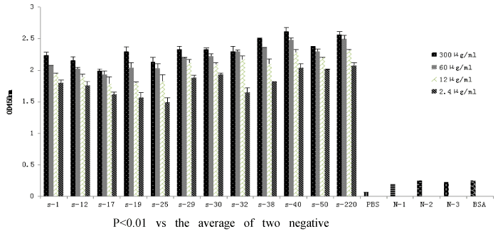
Figure 6:
ELISA analysis of the binding ability of anti-VP2 scFvs to VP2. The plates were coated with different concentrations (300 μg/mL,60 μg/mL,12 μg/mL,2.4 μg/mL) of scFvs, followed by incubation with VP2, chicken egg yolk antibody and secondary antibody. control1 (without VP2 or IBDV strains), control2 (without egg yolk antibody), control3 (without VP2 and egg yolk antibody), control4 (with BSA to replace VP2, or with Newcastle disease virus (NDV) to replace IBDV), and PBS was as background. S denotes scFv, N denotes control. BSA denotes VP2 was replaced by BSA.
Read Full Article HTML DOI: 10.29328/journal.aac.1001001 Cite this Article Read Full Article PDF
More Images
Similar Articles
-
Neutralizing scFv Antibodies against Infectious Bursal Disease Virus Isolated From a Nlpa-Based Bacterial Display LibraryTianhe Li*,Bing Zhou*,Tingqiao Yu,Ning Li,Xiaochen Guo,Tianyuan Zhang,Jingzhuang Zhao,Liming Xu,Siming Li,Lei Ma,Tingting Li,Liangjun Ding,Mingzhe Sun,Deshan Li,Jiechao Yin. Neutralizing scFv Antibodies against Infectious Bursal Disease Virus Isolated From a Nlpa-Based Bacterial Display Library. . 2017 doi: 10.29328/journal.aac.1001001; 1: 001-011
Recently Viewed
-
Chronic endometritis in in vitro fertilization failure patientsAfaf T Elnashar*,Mohamed Sabry. Chronic endometritis in in vitro fertilization failure patients. Clin J Obstet Gynecol. 2020: doi: 10.29328/journal.cjog.1001073; 3: 175-181
-
Relation of Arachnophobia with ABO blood group systemMuhammad Imran Qadir,Sani E Zahra*. Relation of Arachnophobia with ABO blood group system. J Hematol Clin Res. 2019: doi: 10.29328/journal.jhcr.1001011; 3: 050-052
-
Preservation of Haemostasis with Anti-thrombotic Serotonin AntagonismMark IM Noble*,Angela J Drake-Holland. Preservation of Haemostasis with Anti-thrombotic Serotonin Antagonism. J Hematol Clin Res. 2017: doi: 10.29328/journal.jhcr.1001004; 1: 019-025
-
Neutrophil to Lymphocyte Ratio (NLR) in Peripheral Blood: A Novel and Simple Prognostic Predictor of Non-small Cell Lung Cancer (NSCLC)Xiaoli Zhang,Ziyuan Zou,Liyu Fan,Xinjie Xu,Yu Siyuan,Peng Luo*. Neutrophil to Lymphocyte Ratio (NLR) in Peripheral Blood: A Novel and Simple Prognostic Predictor of Non-small Cell Lung Cancer (NSCLC). J Hematol Clin Res. 2017: doi: 10.29328/journal.jhcr.1001002; 1: 011-013
-
Gilbert’s Syndrome Revealed by Hepatotoxicity of ImatinibImen Ben Amor*,Imen Frikha,Moez Medhaffer,Moez Elloumi. Gilbert’s Syndrome Revealed by Hepatotoxicity of Imatinib. Ann Clin Gastroenterol Hepatol. 2025: doi: 10.29328/journal.acgh.1001049; 9: 001-003
Most Viewed
-
Evaluation of Biostimulants Based on Recovered Protein Hydrolysates from Animal By-products as Plant Growth EnhancersH Pérez-Aguilar*, M Lacruz-Asaro, F Arán-Ais. Evaluation of Biostimulants Based on Recovered Protein Hydrolysates from Animal By-products as Plant Growth Enhancers. J Plant Sci Phytopathol. 2023 doi: 10.29328/journal.jpsp.1001104; 7: 042-047
-
Sinonasal Myxoma Extending into the Orbit in a 4-Year Old: A Case PresentationJulian A Purrinos*, Ramzi Younis. Sinonasal Myxoma Extending into the Orbit in a 4-Year Old: A Case Presentation. Arch Case Rep. 2024 doi: 10.29328/journal.acr.1001099; 8: 075-077
-
Feasibility study of magnetic sensing for detecting single-neuron action potentialsDenis Tonini,Kai Wu,Renata Saha,Jian-Ping Wang*. Feasibility study of magnetic sensing for detecting single-neuron action potentials. Ann Biomed Sci Eng. 2022 doi: 10.29328/journal.abse.1001018; 6: 019-029
-
Pediatric Dysgerminoma: Unveiling a Rare Ovarian TumorFaten Limaiem*, Khalil Saffar, Ahmed Halouani. Pediatric Dysgerminoma: Unveiling a Rare Ovarian Tumor. Arch Case Rep. 2024 doi: 10.29328/journal.acr.1001087; 8: 010-013
-
Physical activity can change the physiological and psychological circumstances during COVID-19 pandemic: A narrative reviewKhashayar Maroufi*. Physical activity can change the physiological and psychological circumstances during COVID-19 pandemic: A narrative review. J Sports Med Ther. 2021 doi: 10.29328/journal.jsmt.1001051; 6: 001-007

HSPI: We're glad you're here. Please click "create a new Query" if you are a new visitor to our website and need further information from us.
If you are already a member of our network and need to keep track of any developments regarding a question you have already submitted, click "take me to my Query."


